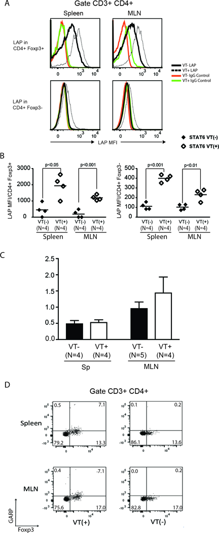
Figure 8. Continuous expression of active STAT6 in T cells is sufficient to stimulate the expression of pro-TGFβ.

(A) Mean fluorescence intensity of LAP staining on Foxp3+ CD4+ and Foxp3- CD4+ T cells from spleen and MLN of STAT6 VT (STAT6 VT+; white histograms) mice and WT (STAT6 VT-) littermates (gray histograms). (B) Cumulative data showing mean±SD from several experiments (n: the number of independent experiment. Each dot represents a single experiment with bars representing mean values from multiple experiments; p value as indicated in each panel). (C) TGFβ mRNA content in purified splenic or MLN CD4 T cells from STAT6 VT- (Black rectangles) or STAT6 VT+ (white rectangles) mice were displayed as the ratio of TGFβ mRNA to HPRT mRNA. (N: number of independent measurements; p: not significant between STAT6 VT- and STAT6 VT+ groups). (D) Spleen and MLN cells from uninfected and helminth-infected STAT6 VT+ (VT(+)) or STAT6 VT- (VT(−)) mice were stained for CD3, CD4, Foxp3 and GARP. Cells were gated on CD3+ CD4+ T cells. Numbers represent the percentage of events in each quadrant. Data are representative example from three samples for each set.
