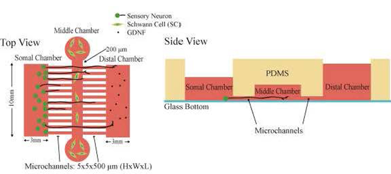
Figure 3: Schematics of the microfluidic device.
A) Top and B) side views of the microfluidic device. Both Somal and Distal chambers have a surface of 10 mm×3 mm (schematics are not drawn to scale). Middle chamber reservoirs are 5mm in diameter and the chamber dimensions are 100 mm × 200 μm × 100 μm (L×W×H). The two sets of microchannels dimensions are 5 × 5 × 500 (L×W×H) μm. Side view shows different volumes applied to each chamber. The volume applied to the middle chamber is the volume applied to the two circular reservoirs.

