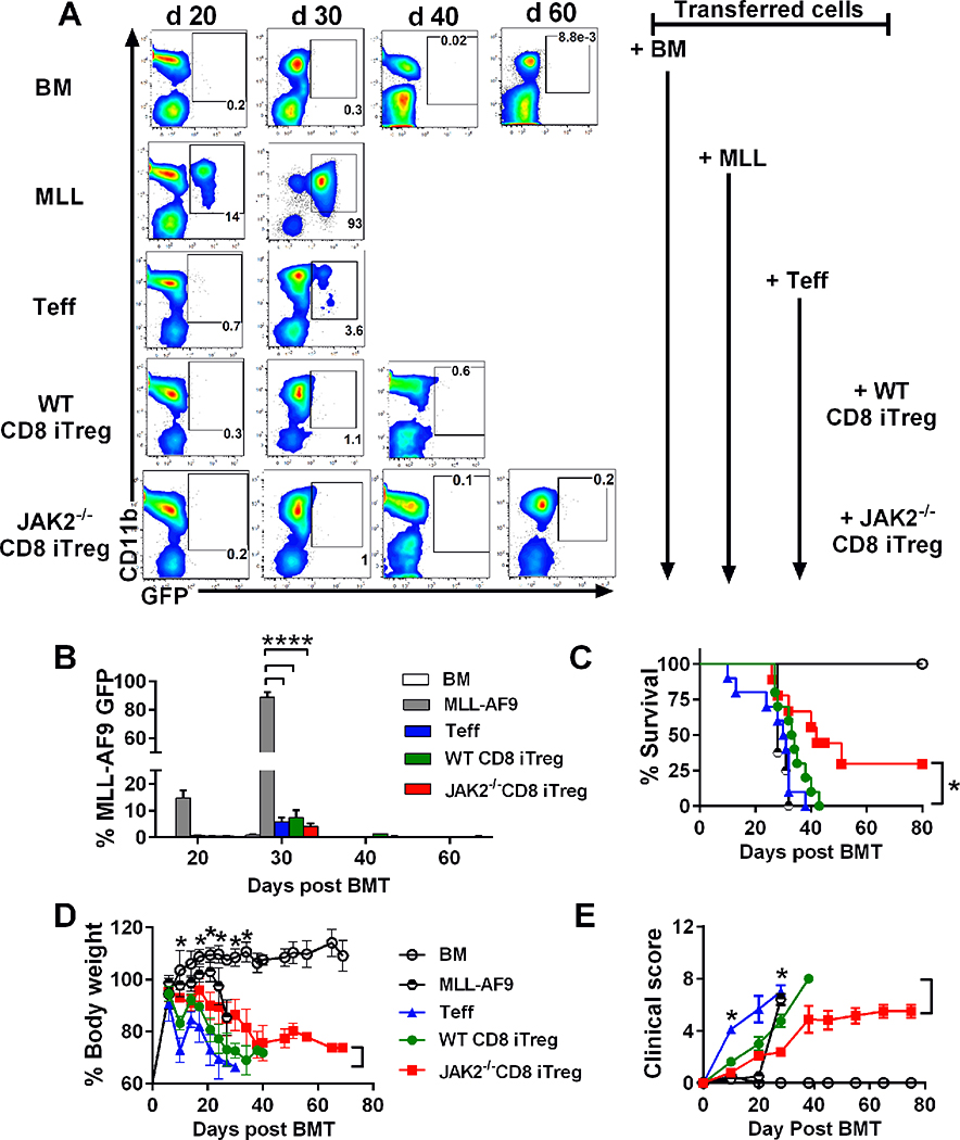
FIGURE 7. Effects of CD8+ iTregs on GVH and GVL responses.
Lethally irradiated BALB/c mice were adoptively transferred with 1×10⁶ CD8+ iTregs, 5×10⁶ WT-TCD BM, and 2×10⁴ MLL-AF9. Three days later, 0.7×10⁶ CD25-depleted T-cells were i.v injected to induced aGVHD. (A) % MLL-AF9 in recipient peripheral blood were analyzed with CD11b+GFP+, and representative FACS plots on day 20, 30, 40 and 60 posts BMT were shown. (B) Bar graph showed quantified %GFP+ MLL-AF9 in blood at the indicated time point. Survival rate (C), body weight loss (D), and GVHD clinical signs (E) of recipient mice were monitored until day 60. Data are combined from 2 independent experiments (n=8–10/group). Log-rank (Mantel-Cox) test was used to compare the survival data. Student’s t-test was used to compare % MLL, body weight loss and GVHD clinical sign data. *p ≤ 0.05, **p ≤ 0.01 and, ****p ≤ 0.0001. Data represent the mean ± SEM.

