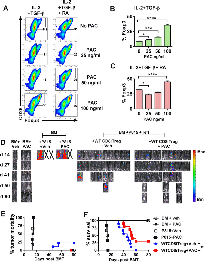
FIGURE 8. Impact of Pacritinib and iTregs in GVHD and tumor relapse.
Allogeneic WT CD8+ iTregs were generated in vitro in the presence or absence of Pacritinib (PAC) at the indicated concentration. After 5 days incubation, Foxp3 expression was analyzed by flow cytometry. (A-C) FACS plots and graphs show %Foxp3 expression on CD8+ cells (n=3/group) under indicated conditions. Student’s t-test was used to compare the data. *p ≤ 0.05, ***p ≤ 0.001 and, ****p ≤ 0.0001. Data represent the mean ± SEM. (D-F) Lethally irradiated BDF1 mice were adoptively transferred with 2×10⁶ WT CD8+ iTregs, 5×10⁶ WT-TCD BM, and 2–5 ×10³ P815 mastocytoma. Three days later, 3 ×10⁶ CD25-depleted T-cells were i.v injected. Pacritinib 100 mg/kg/day or methylcellulose vehicle was given by oral gavage daily for 4 weeks starting on day 0 of BMT. Recipients were monitored for tumor burden (D) tumor mortality (E) and survival (F) until day 80. Data are combined from 2 independent experiments (n=9–10/group). *p ≤ 0.05. Log-rank (Mantel-Cox) test was used to compare the tumor mortality and survival.

