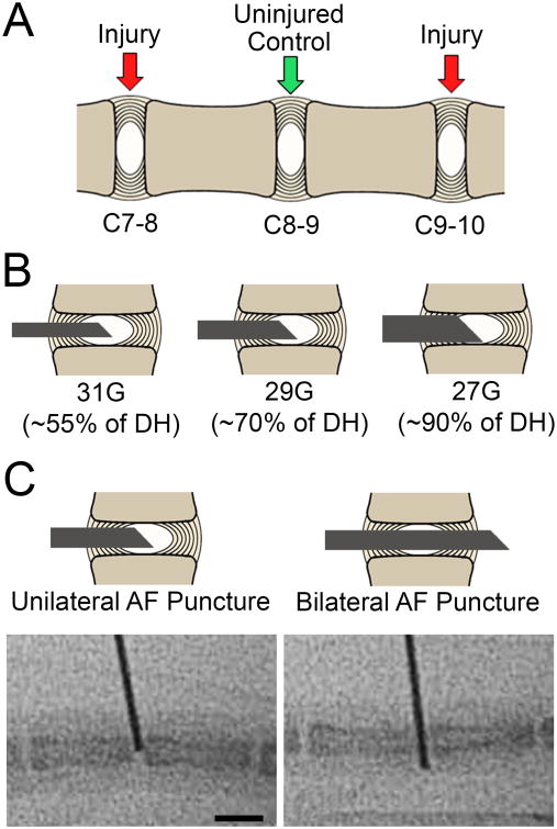
Figure 1.
A. Schematic representation of the mouse caudal spine showing the three experimental disc levels. B. Schematic representations of the three needle sizes used for disc injury. C. Schematic representations and corresponding intraoperative fluoroscopy images (below), showing the two injury types investigated for each needle size (unilateral or bilateral AF puncture). Scale = 5 mm.

