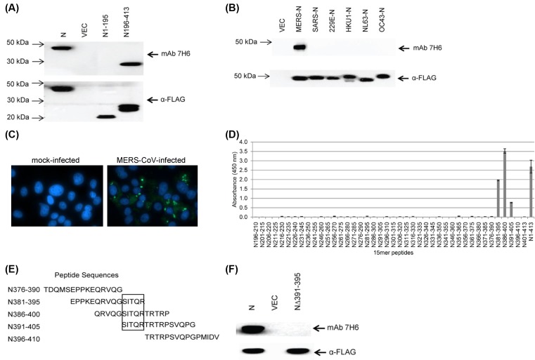
Figure 1. Characterization of mAb 7H6.
(A) 293FT cells were transiently transfected with empty vector, FLAG-tagged full-length MERS-N, and its N- and C-terminal fragments. Western blot analysis was performed using mAb 7H6 and anti-FLAG antibody. (B) 293FT cells were transiently transfected with empty vector or FLAG-tagged N of different HCoVs. Western blot analysis was performed using mAb 7H6 or anti-FLAG antibody. (C) Vero E6 cells were mock infected or infected with MERS-CoV (multiplicity of infection of 1) and stained with mAb 7H6 at 2 days post-infection. (D) 15-mer peptides with ten amino acids overlapping sequences of the C-terminal fragment of MERS-N were generated and their binding to mAb 7H6 was analyzed by ELISA. Three independent experiments were performed and a representative data is shown. The results are mean values with error bars showing S.D. of triplicate wells. (E) Peptides between N376-410 were aligned and the minimal binding sequence was mapped (denoted in a box). (F) 293FT cells were transiently transfected with empty vector, FLAG-tagged MERS-N and mutant MERS-NΔ391-395. Western blot analysis was performed using mAb 7H6 and anti-FLAG antibody.

