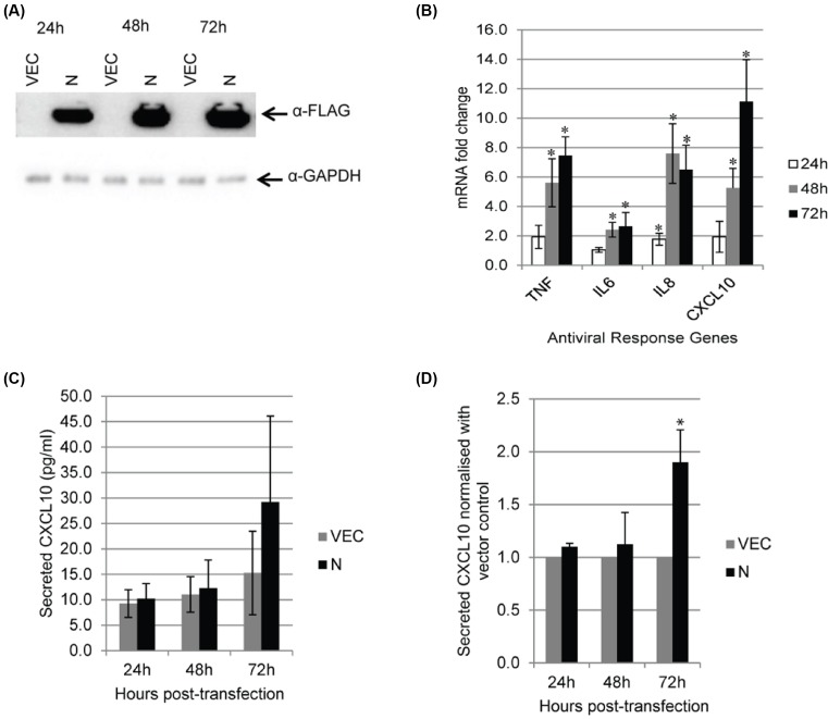
Figure 3. Expression of the four selected antiviral genes in 293FT cells.
(A) 293FT cells were transiently transfected with empty vector and FLAG-tagged MERS-N. Western blot analysis was performed using anti-FLAG antibody. (B) RNA transcripts from transiently transfected 293FT cells were assayed by RT-qPCR with TaqMan probes to analyze the mRNA fold changes of TNF, IL6, IL8, and CXCL10. mRNA expression of genes was first normalized against GAPDH and subsequently, MERS-N up-regulation was normalized against levels in vector-transfected cells. (C) Cell supernatants were harvested and CXCL10 secretion was evaluated using ELISA. (D) The secretion of CXCL10 by MERS-N was normalized against the vector. The results of these experiments were expressed as mean ± S.D. (error bars) of five independent experiments. Asterisk (*) indicates statistical significance of P<0.05 when compared with vector-transfected cells at the respective time-points.

