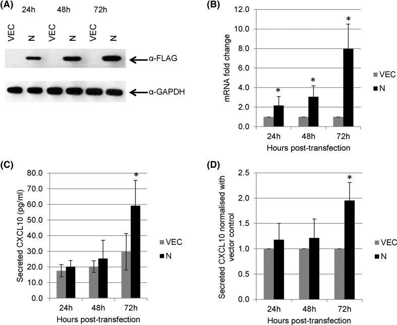
Figure 4. Expression of CXCL10 in A549 cells.
(A) A549 cells were transiently transfected with empty vector and FLAG-tagged MERS-N. Western blot analysis was performed using anti-FLAG antibody. (B) RNA transcripts from transiently transfected A549 cells were assayed by RT-qPCR with TaqMan probes to analyze the mRNA fold changes for the expression of CXCL10. mRNA expression of CXCL10 was first normalized against GAPDH and then normalized against levels in vector-transfected cells. (C) Cell supernatants were harvested from the transiently transfected cells, and CXCL10 secretion was evaluated using ELISA. (D) The secretion of CXCL10 of cells overexpressing MERS-N was normalized against the vector. The results of these experiments were expressed as mean ± S.D. (error bars) of six independent experiments in triplicates. The asterisk (*) indicates statistical significance of P<0.05 when MERS-N is compared with vector control.

