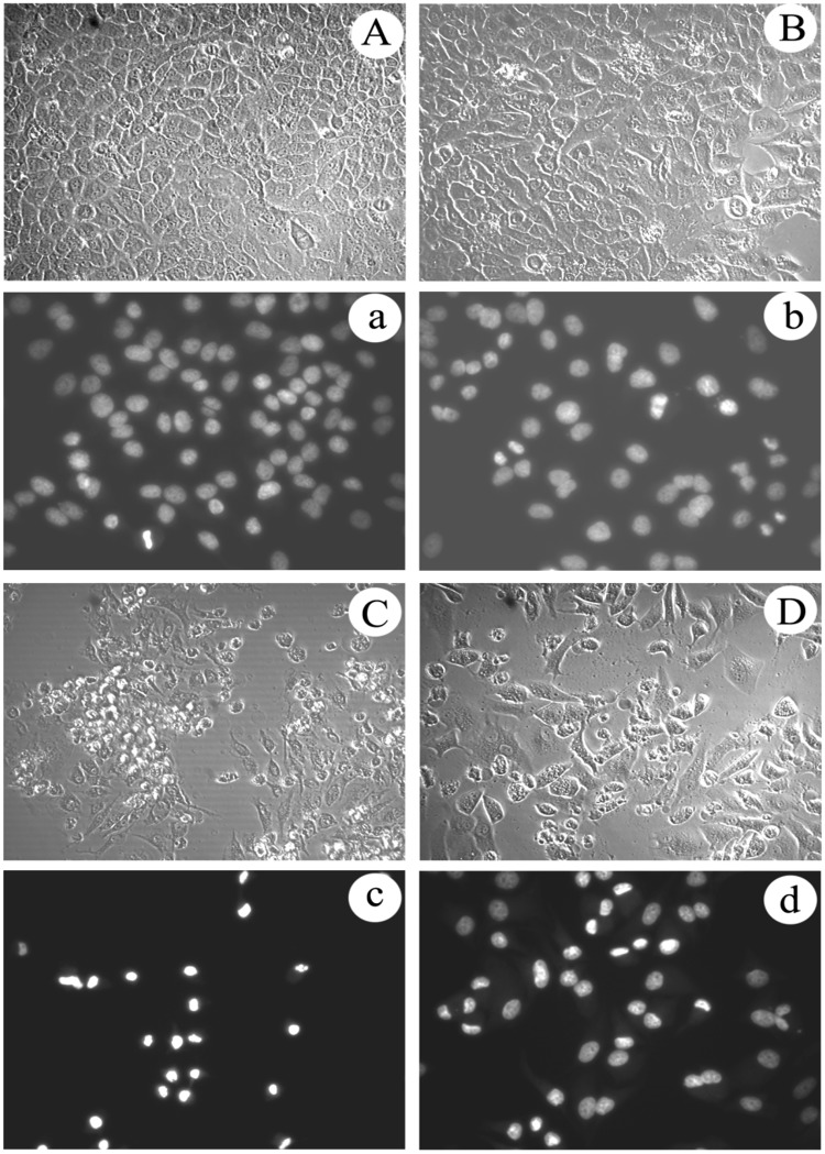
Figure 9.
Analysis of proliferation and cell morphological change in Hela229 cells treated with matrine derivatives 1 and 2 for 48 h. Photographs marked with capital letters were obtained from IPCM (inverted phase contrast microscopy), with small letters from FM (fluorescence microscopy); magnification was 200X . (A) Morphological characteristics of cell in control; (a) Morphological characteristics of cell nucleus in control; (B) Morphological changes of cell induced by matrine; (b) Nucleus morphological changes of cell induced by matrine; (C) Morphological changes of cell induced by matrine derivative 1; (c) Nucleus morphological changes of cell induced by matrine derivative 1; (D) Morphological changes of cell induced by matrine derivative 2; (d) Nucleus morphological changes of cell induced by matrine derivative 2.

