
Figure 5.
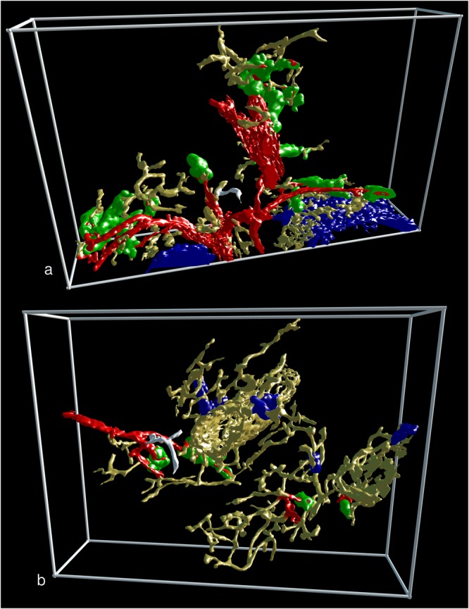
Two non-sheathed capillaries arising from an arterial vessel proximal to a capillary sheath. The non-sheathed capillaries are marked in white colour. All non-connected structures were removed. (a) Part of R3 seen from the last section in the series. (b) Part of R8 seen from the first section in the series. (a) and (b) show staining for CD34 plus SMA in yellow, for post-arteriolar CD271+ capillary sheaths in green and for capillary sheaths of undetectable location in blue. In (a) the blue structure in the lower right part corresponds to CD271+ FDCs in follicles. The direct connections of arterial vessels to green sheaths were manually marked in red. Length of the horizontal part of the bounding box = 882 µm in (a) and 709 µm in (b).