Fig. 6.
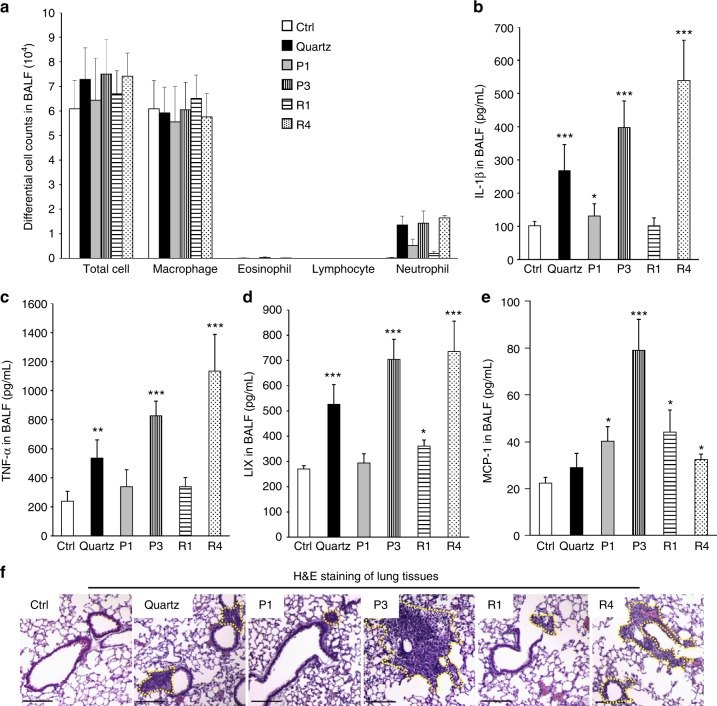
Inflammatory effects and cell migration in Fe2O3-treated animal lungs. a Differential cell counts, b IL-1β, c TNF-α, d LIX, and e MCP-1 productions in BALF, and f H&E staining of lung sections from Fe2O3-treated mice. Selected Fe2O3 nanoplates (P1, P3) and nanorods (R1, R4) were oropharyngeally administrated at 2 mg/kg (n = 6 mice in each group), whereas that animals received 5 mg/kg quartz exposure were used as positive control. After 40 h, animals were killed to collect BALF for differential cell counting as well as cytokine measurements, including IL-1β, LIX, TNF-α, and MCP-1. Data are shown as mean ± SD from four independent replicates. *p < 0.05, **p < 0.01, ***p < 0.001 compared with vehicle control (two-tailed Student’s t-test). The dashed lines in H&E staining images show the immune cell recruitments and the scale bar represents 200 μm

