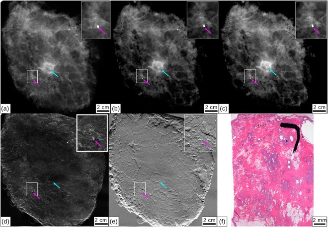
Figure 3.
Clinical mammography and monochromatic absorption-contrast and grating-based multimodal mammography for specimen III. (a) Clinical ex-vivo absorption-contrast mammography (cevAC-Mx). (b) Monochromatic absorption-contrast mammography (mAC-Mx). (c) Monochromatic grating-based absorption-contrast mammography (mgbAC-Mx), (d) dark-field mammography (mgbDFC-Mx) and (e) differential phase-contrast mammography (mgbDPC-Mx). Inlets show a calcification cluster that had previously been marked. The clip marker is highlighted with a magenta arrow, a light blue arrow indicates the mamilla. All images were scaled for maximum detail visibility. (f) Histopathology of the mastectomy sample showing microcalcifications.

