Figure 1.
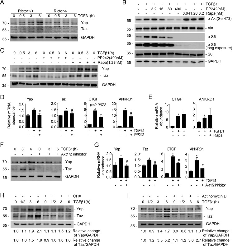
Blockade of Rictor/mTORC2 signaling reduces TGFβ1-induced Yap/Taz expression. A, the primary cultured fibroblasts generated from the kidneys of Rictorfl/fl mice were infected with adeno-Cre virus. After 48 h, the fibroblasts were serum-starved overnight and then treated with TGFβ1 (2 ng/ml). Western blot assays show the abundance for Yap/Taz. GAPDH was probed to show the equal loading of the samples. B, Western blot analyses showing the abundance of p-Akt (Ser-473) and p-S6 in NRK-49F cells after PP242 or rapamycin treatment. C, NRK-49F cells were pretreated with PP242 (400 nm) and rapamycin (1.28 nm) for 30 min, followed by TGFβ1 (2 ng/ml) administration for different durations. The Western blot assay demonstrated that PP242 but not rapamycin could inhibit TGFβ1-induced Yap/Taz expression. D, real-time PCR analysis showing the mRNA abundance for Yap, Taz, CTGF, and ANKRD1 in NRK-49F cells. *, p < 0.05 compared with control cells (n = 3–4); #, p < 0.05 compared with cells treated with TGFβ1 (n = 3–4). E, real-time PCR analysis showing the mRNA abundance for CTGF and ANKRD1 in NRK-49F cells. *, p < 0.05 compared with control cells (n = 3). F, Western blot analysis showing that Akt1/2 inhibitor could suppress TGFβ1-induced Yap and Taz expression. G, real-time PCR analysis showing the mRNA abundance for Yap, Taz, CTGF, and ANKRD1 in NRK-49F cells. *, p < 0.05 compared with control cells (n = 3–4); #, p < 0.05 compared with cells treated with TGFβ1 (n = 3–4). H and I, NRK-49F cells were treated with cycloheximide (50 μg/ml) or actinomycin D (0.1 μm), followed by TGFβ1 (2 ng/ml) administration. A Western blot assay shows the abundance for Yap and Taz. GAPDH was probed to show the equal loading of the samples. Error bars, S.E.

