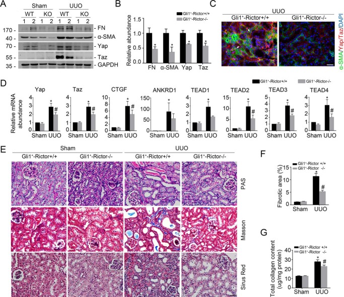
Figure 4.
Ablation of Rictor in fibroblasts diminishes Yap/Taz activation and UUO nephropathy in mice. Mice were performed with UUO and sacrificed at day 7 after surgery. A and B, Western blot assay (A) and quantitative analysis (B) showing FN, α-SMA, Yap, and Taz protein abundance in the Sham and UUO kidneys from Gli1+-Rictor+/+ and Gli1+-Rictor−/− mice, respectively. Numbers indicate individual animals within each group. *, p < 0.05 compared with Gli1+-Rictor+/+ littermates after UUO (n = 4). C, representative images showing the lesser induction of Yap/Taz protein in the kidney myofibroblasts from the Gli1+-Rictor−/− mice. White arrows, Yap/Taz-positive myofibroblasts. Scale bar, 20 μm. D, real-time PCR analysis showing the mRNA abundance for Yap, Taz, CTGF, ANKRD1, and TEAD1–4 from Sham and UUO kidneys. *, p < 0.05 versus Sham control (n = 5–6); #, p < 0.05 versus Rictor+/+ littermates after UUO (n = 5–6). E, representative images of periodic acid–Schiff (PAS), Masson, and Sirius red staining in the kidneys from different groups as indicated. Scale bar, 20 μm. F and G, graphic presentation showing the fibrotic area and total collagen content in kidney tissues among groups as indicated. *, p < 0.05 compared with Sham control (n = 4–5); #, p < 0.05 compared with UUO kidneys from control littermates (n = 4–5). Error bars, S.E.

