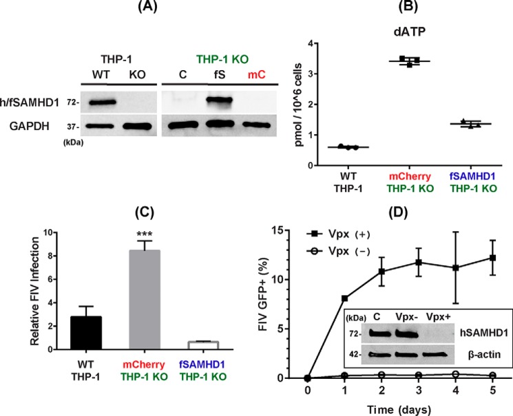
Figure 2.
Cellular dNTP reduction activity and antiviral activity of fSAMHD1 in nondividing THP-1 macrophages. A, expression of fSAMHD1 in hSAMHD1 KO THP-1 cells. Left, human SAMHD1 expression in the PMA-treated parental THP-1 macrophages (WT) was previously abolished by CRISPR/Cas9 (KO) (33). hSAMHD1 expression was detected by Western blotting with anti-SAMHD1 antibody in the differentiated THP-1 cells. Cellular GAPDH protein was used as a loading control. Right, hSAMHD1 KO THP-1 cells (THP-1 KO) were transduced with lentiviral vector co-expressing both HA-tagged fSAMHD1 (fS) and mCherry protein or lentiviral vector expressing only mCherry protein (mC), and the mCherry+ cells were FACS-sorted. The sorted mCherry+ THP-1 were then differentiated to macrophages by PMA for 7 days and analyzed for the fSAMHD1 expression (fS) by Western blotting with HA antibody. GAPDH was used as a loading control. C, untransduced SAMHD1 KO THP-1 control cells. B, cellular dNTP reduction by fSAMHD1 expression in nondividing hSAMHD1 KO THP-1 macrophages. The parental THP-1 cells expressing hSAMHD1 (WT THP-1), hSAMHD1 KO THP-1 cells expressing only mCherry protein (mCherry THP-1 KO), and hSAMHD1 KO THP-1 cells expressing both fSAMHD1 and mCherry protein (fSAMHD1 THP-1 KO) were differentiated by PMA for 7 days, and the dNTP levels in these cells were determined by the RT-based dNTP assay. The total dNTP amounts were normalized by 1 × 10⁶ cells. dATP levels are shown in this figure, and other three dNTP data are in Fig. S2. C, FIV restriction by fSAMHD1 in nondividing hSAMHD1 KO THP-1 macrophages. The parental THP-1 cells expressing hSAMHD1 (WT THP-1), hSAMHD1 KO THP-1 cells expressing only mCherry protein (mCherry THP-1 KO), and hSAMHD1 KO THP-1 cells expressing HA-tagged fSAMHD1 were differentiated by PMA, and transduced with an equal amount of FIV-GFP vector with error bars representing S.D. ***, p ≤ 0.001, using two-tailed Student's t test. The -fold changes of the GFP+ cells compared with the untransduced THP-1 negative background control cells (ratio = 1) in triplicates were plotted. The transduction efficiencies of the vectors used (% GFP ± S.D.) were 1.187 ± 0.384, 3.567 ± 0.361, and 0.813 ± 0.101 for WT THP-1, mCherry THP-1 KO, and fSAMHD1 THP-1 KO, respectively. D, FIV restriction by hSAMHD1 in human primary monocyte-derived macrophages. Human primary monocyte-derived macrophages prepared from four healthy donors were pretreated with VLPs with (Vpx+) or without Vpx (Vpx−) for 12 h, and then transduced with an equal amount of FIV-GFP vector. The transduced cells were collected every day for 5 days, and the percent of the GFP+ cells in triplicates was determined by FACS. Insert shows Western blotting conducted to confirm the degradation of hSAMHD1 by Vpx in human primary macrophages: β-actin was used as a loading control, and antiSAMHD1 antibody was used for detecting hSAMHD1 in the human primary macrophages. C, no VLP pretreatment control macrophages.

