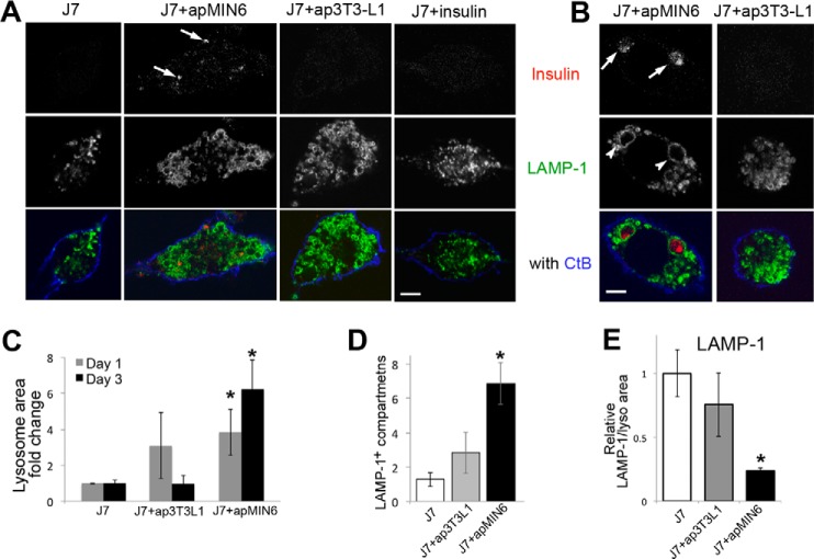
Figure 2.
Phagocytosis of apMIN6 cells leads to insulin accumulation and enlarged lysosomes. A and B, incubation with apMIN6, but not ap3T3-L1 or monomeric insulin, induced insulin accumulation, and lysosome swelling. One day after efferocytosis, insulin was detected in LAMP-1 positive lysosomes indicated by the arrows (A). By day 3, enlarged lysosomes (arrowheads) were found in J7 cells incubated with apMIN6 (B) by confocal microscopy. Scale bars, 5 μm. C, two-dimensional lysosome area was measured by manually tracing the lysosomes outlined by LAMP-1. D, lysosome outlines were determined by LAMP-1, and those with a diameter greater than 2 μm were counted. E, LAMP-1 fluorescence intensity was measured and normalized to lysosome area. C–E, data are presented as fold-change relative to J7 cells alone. n = 48 lysosomes from 9 cells. *, p < 0.05 versus J7 alone.

