Figure 5.
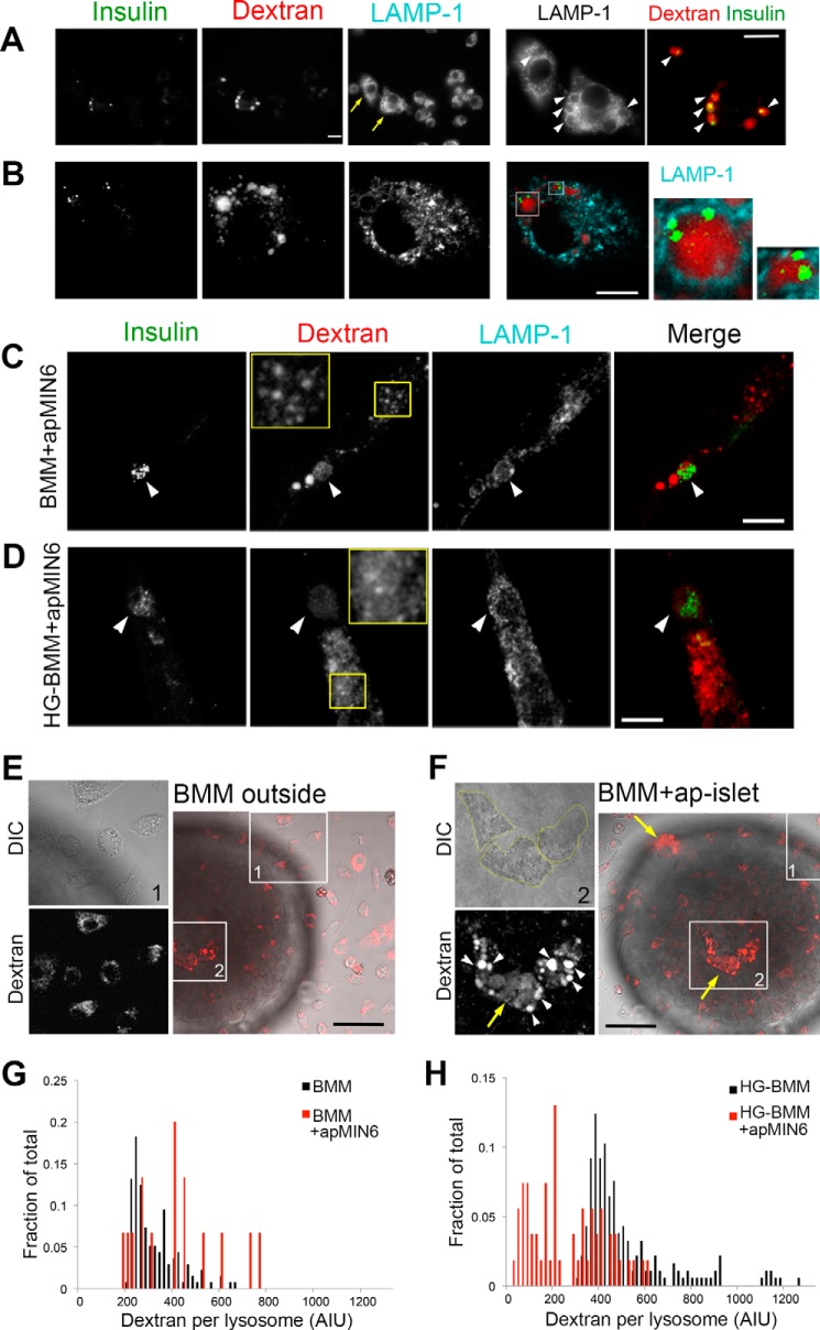
High glucose induces lysosomal permeabilization in phagocytic macrophages. A and B, dextran and insulin aggregates co-exist in intact lysosomes. J7 cells (A) or BMMs (B) were loaded with lysine-fixable rhodamine dextran (red) after phagocytosis of apMIN6 cells, fixed, and labeled with insulin (green) and LAMP-1 (cyan) antibodies before being imaged by wide-field (A) and confocal (B) microscopy. The yellow arrows in A show two cells with insulin aggregates in enlarged lysosomes filled with dextran (arrowheads). The two cells are expanded in the bottom panels in A. Single plane images are shown in B. Two lysosomes are expanded to the right, which show co-existence of dextran and insulin. C and D, BMMs were incubated with apMIN6 cells and cultured in either normal medium (C, BMMs+apMIN6) or high glucose medium for 3 days (D, HG-BMMs+apMIN6). They were then loaded with lysine-fixable rhodamine-dextran and labeled with insulin and LAMP-1 antibodies. Shown here are summation projection images by confocal microscopy. The yellow boxes show a region of the cell in which the dextran shows punctate staining in the lysosomes (C) or additional diffuse staining in the cytoplasm (D). The merged images show insulin in green and dextran in red. Scale bar, 10 μm (A–D). E and F, HG-BMMs loaded with rhodamine-dextran were added to apoptotic islets and imaged live by confocal microscopy. Images in E and F focused on the BMMs that were outside (box 1) and inside (box 2) the same islet, respectively. White arrowheads show enlarged lysosomes and yellow arrows show diffuse dextran in the cytoplasm (F). Scale bars, 50 μm. G and H, quantification of dextran intensity in lysosomes of BMMs (G) or HG-BMMs (H) with or without phagocytosis of apMIN6 cells. Lysosomes of all cells under the same condition were pooled from one experiment; the results are representative of three experiments.

