Figure 7.
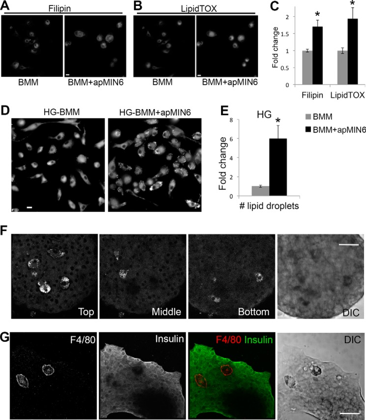
Lipid droplet formation in phagocytic HG-BMMs. A–C, BMMs cultured in normal growth medium showed increased cholesterol (labeled by filipin in A) and neutral lipid (labeled by LipidTOX in B) accumulation after β-cell efferocytosis. Total fluorescence intensity per cell was quantified in C from 10 imaging fields. D and E, BMMs cultured in HG medium showed increased lipid droplet formation (labeled by LipidTOX) after β-cell efferocytosis. Number of lipid droplets per cell was quantified in E from 9 imaging fields. A–E, scale bars, 10 μm; *, p < 0.05 versus BMM alone. F, isolated islets were cultured in HG, labeled with LipidTOX, and imaged by confocal microscopy. Three images of the same islet show islet macrophages present in different focal planes. G, isolated islets were cultured in HG, labeled with F4/80 and insulin antibodies, and imaged by confocal microscopy. DIC, differential interference contrast. Scale bars, 50 μm.

