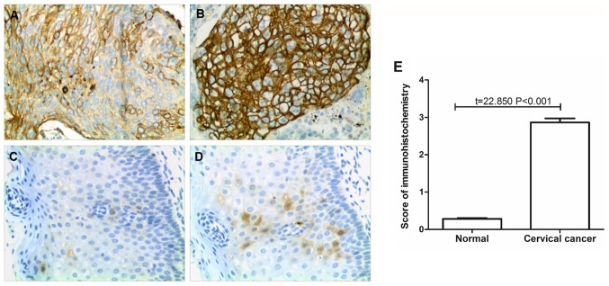
Figure 1.
Detection of TF expression by immunohistochemistry. (A) Low TF expression in cervical cancer tissue. (B) High TF expression in cervical cancer tissue. (C) Negative TF expression in adjacent normal tissue. (D) Low TF expression in adjacent normal tissue. Magnification, ×200. (E) Statistical analysis of the immunohistochemistry results. TF, tissue factor.

