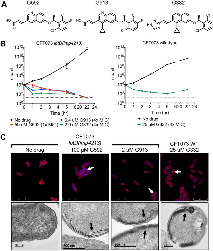
FIG 2.
Small-molecule inhibitors of E. coli MsbA. (A) Chemical structures of the three quinoline inhibitors described in this paper. (B) Time-kill assay of CFT073 lptD(imp4213) and CFT073 with MsbA inhibitors. Viable cells were measured by plating on LB agar at 0, 1, 2, 3, 5, and 22 h after addition of compound. (C) Confocal microscopy and TEM of CFT073 and CFT073 lptD(imp4213) treated with the indicated concentration of inhibitor for 3 h. For confocal microscopy, membranes were stained with Nile red, and DNA was stained with DAPI (blue). Arrows in confocal and TEM images indicate sites of excess membrane accumulation. Untreated CFT073 cells looked identical to untreated CFT073 lptD(imp4213) cells (data not shown).

