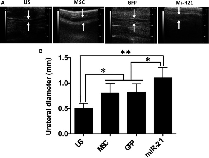
Figure 5.

Micro‐ultrasound observation and detection of urethral diameter. A, Representative micro‐ultrasound images of rat penile urethras from different groups, and measurement of urethral diameter accordingly 4 wk after injections (n = 6), *P < 0.05, **P < 0.01
