Figure 3.

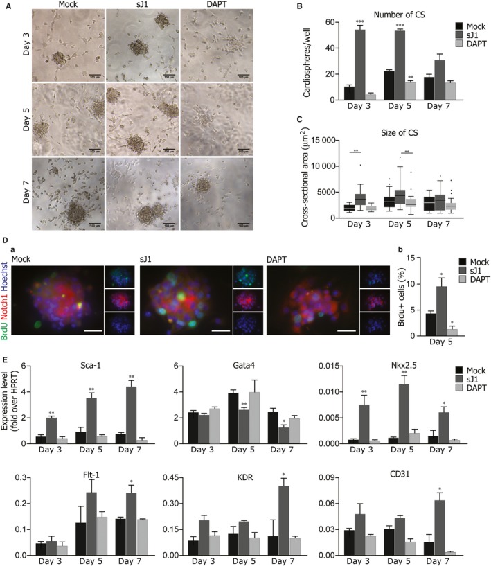
Stimulation of the Notch pathway enhances cardiosphere growth and induces a cardiovascular differentiation programme. Cardiosphere‐forming cells were treated either with the soluble form of Jagged1 (sJ1) or with DAPT for 24 h before the analysis after 3, 5 and 7 days of in vitro culture A, Representative bright field images of cardiospheres, treated either with 20x sJ1 or DAPT, after 3, 5 and 7 days of in vitro culture. Scale bar: 100 μm. B, Quantification of the number cardiospheres treated either with 20x sJ1 or DAPT, after 3, 5 and 7 days of in vitro culture. Shown are mean ± SEM of at least 3 experimental replicates ***P < 0.001; **P < 0.01. C, Distribution of the cross‐sectional areas of the cardiospheres, treated either with 20x sJ1 or DAPT, after 3, 5 and 7 days of in vitro culture. Shown are Tukey boxplots, **P < 0.01. D, (a) Representative images of BrdU+ cells in cardiospheres after 5 days of in vitro culture in the presence of 20X sJ1 or DAPT. BrdU (green), Notch1 (red), Nuclei (blue). Scale bar 40 μm. (b) Quantification of BrdU+ cells upon sJ1 or DAPT treatment. Shown are mean ± SEM of at least 3 experimental replicates. **P < 0.01: *P < 0.05. E, Transcription levels of indicated genes in cardiospheres analysed at day 3, 5 and 7 of in vitro culture, in the presence of either 20x sJ1 or DAPT. Data are expressed to cellular HPRT mRNA levels. Shown are the mean ± SEM of at least 3 independent experiments. **P < 0.01: *P < 0.05
