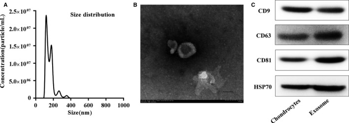
Figure 1.

Characterization of chondrocytes‐derived exosomes. A, Particle size distribution of exosomes measured by Nanosight. B, Morphology of exosomes observed by transmission electron microscopy (TEM). Scale bar 100 nm. C, Exosome surface markers (CD9, CD63, CD81, and Hsp70) measured using western blotting. The cartilage exosomes were obtained by chondrocytes culture supernatants via ultracentrifugation. This experiment was repeated independently three times and representative results are shown
