
Figure 3. CaMKII-dependent and PKA-dependent regulation of basal INaL under AP-clamp.
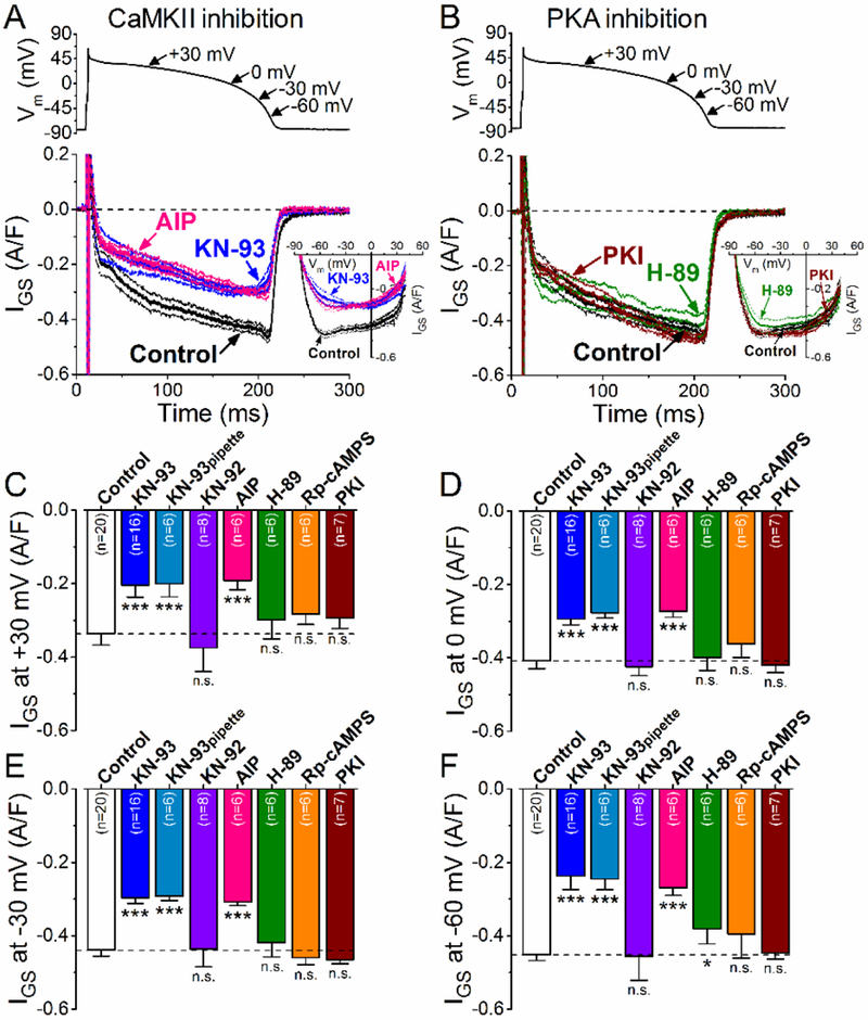
(A) GS-458967-sensitive current (IGs) traces (mean±SEM) under AP-clamp in control and after CaMKII inhibition using KN-93 (1 pM) or AIP (1 μM). The physiological intracellular Ca2+ cycling was preserved, and 2 Hz steady-state pacing was applied. I-V trajectories of IGS under AP-clamp are shown in inset. (B ) IGS traces (mean±SEM) under AP-clamp in control and after PKA inhibition using H-89 (1 μM) or PKI (1 μM). I-V relationships are shown in the inset. (C-E) IGS density at different voltages during AP repolarization. CaMKII inhibition with KN-93 or AIP significantly decreased INaL at all voltages, whereas KN-92 (1 μM) had no effect on IGS. The decrease in INaL was most prominent at −60 mV which may reflect the window component of INaL. On the contrary, PKA inhibition using PKI or Rp-cAMPS (100 μM) had no effect on IGS, whereas H-89 may exhibit some off-target effect at −60 mV. Columns and bars represent mean±SEM. Asterisks denote significant difference using two-way ANOVA with Bonferroni posttest. *p<0.05, **p<0.01, ***p<0.001.