Fig. 5.
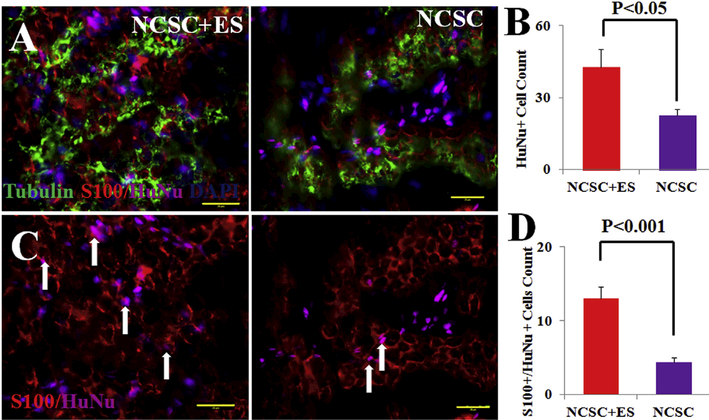
At 12 weeks, (A) hNCSC localization and differentiation were confirmed as evident by immunostaining of HuNu (magenta), S-100 (red), and Tubulin (green) in NCSC + ES and NCSC transplanted nerves; (B) qualification of HuNu positive cells showed that NCSC + ES has superior survivability than NCSC alone. (C) co-staining human cell nuclei (HuNu, magenta) and S-100 (red) expression indicated donor human cell-derived S100 positive cells (arrows) 12 weeks after NCSC transplantation; (D) qualification ofboth S100 and HuNu positive cells explained that hNCSCs were able to survive or differentiate into peripheral nerve support cells, and ES further promoted hNCSCs survival and differentiation in vivo. Blue = nuclear staining with DAPI. Scale bar represents 20 μm. (For interpretation of the references to colour in this figure legend, the reader is referred to the Web version of this article.)

