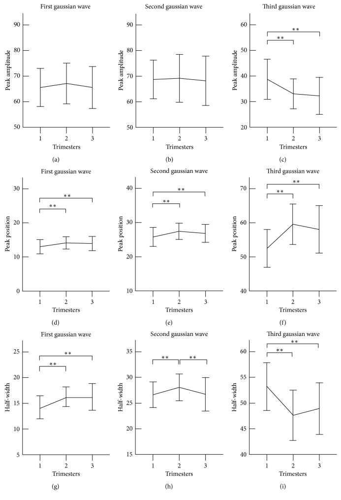
Figure 3.
Gaussian modelling characteristics of the radial pulse measured at the three trimesters of healthy pregnant women. Their means ± SDs are given. (a–c) show peak amplitude changes of the first, second, and third Gaussian waves (H1, H2, H3) at the three trimesters; (d–f) show changes of peak time position of the first, second, and third Gaussian waves (N1, N2, N3) at the three trimesters; (g–i) shows changes of half-width of the first, second, and third Gaussian waves (W1, W2, W3) at the three trimesters. ∗∗ represents p < 0.01.

