Figure 1.

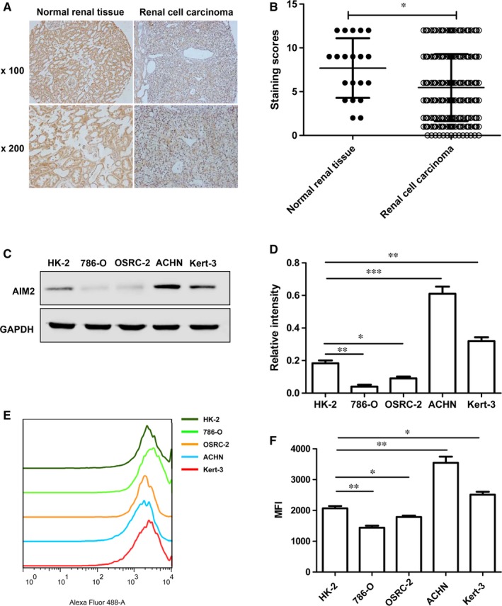
AIM2 expression was decreased in human renal cell carcinoma (RCC) and renal cell lines. (A). This represents immunohistochemical staining of AIM2 in RCC and normal renal tissues, top panel,×100, bottom panel, ×200. (B). Staining scores of AIM2 were evaluated in (A), the immunohistochemical staining data were available from 20 normal renal tissues and 298 RCC. (C). AIM2 expression was evaluated in both 786‐O and OSRC‐2 cell lines by Western blot. (D). The relative values were estimated in the band intensity of each band normalized by GAPDH. (E). The expression levels of AIM2 were detected by flowcytometry in HK‐2, 786‐O, OSCR‐2, ACHN and Kert‐3 cells. (F). The data shown as statistical analysis of the mean fluorescence intensity in (E). Data represent the means of three independent experiments. Data are shown as means ± SD. The different significance was set at *P < 0.05, **P < 0.01 and ***P < 0.001
