Figure 2.

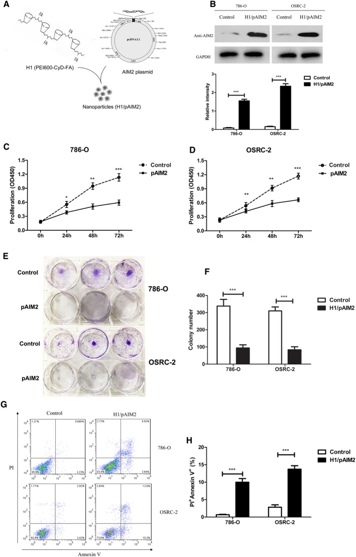
H1/AIM2 nanoparticles inhibited cell proliferation and promoted cell apoptosis in renal cancer cells. (A). A schematic of H1/pAIM2 nanoparticle formation. (B). Forty‐eight hours after H1/pAIM2 transfection, AIM2 expression was evaluated by Western blot in both 786‐O and OSRC‐2 cell lines. (B). The values of the band intensity below the figure represent the densitometric estimation of each band normalized by GAPDH. (C and D). CCK‐8 cell proliferation assay was performed after H1/AIM2‐treated in 786‐O and OSRC‐2. (E and F). The colony formation capability of H1/pAIM2 or control‐treated renal cancer cells were detected at day 7. (G) H1/pAIM2 The cell apoptosis was detected by flow cytometry in 786‐O and OSRC‐2. (H). Statistical histograms of the percentage of apoptotic cells among H1/pAIM2‐, and control‐treated 786‐O and OSRC‐2 cells, respectively. All experiments were carried out in triplicate. Data are shown as means ± SD. The different significance was set at *P < 0.05, **P < 0.01 and ***P < 0.001
