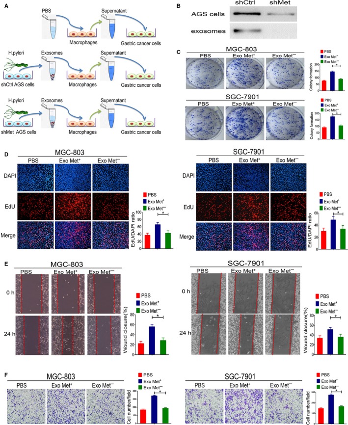
Figure 4.

Exosomal mesenchymal‐epithelial transition factor (MET) mediates pro‐tumorigenic effects in macrophages. A, Schematic description of the experimental design. B, Suppression efficiency of MET shRNA in AGS cells or AGS cell‐derived exosomes was detected by Western blotting. C, Colony formation was evaluated based on GC cell proliferation. D, Representative images and quantification of DNA synthesis (EdU assay) in MGC‐803 and SGC‐7901 cells co‐cultured with supernatant from macrophages stimulated with PBS, MET+ exosomes, or MET− exosomes. Scale bars represent 50 μm. E, Cell migration was assessed using a wound‐healing assay. F, A transwell assay was performed to assess GC cell invasion. Data represent at least three experiments performed in triplicate. *P < 0.05
