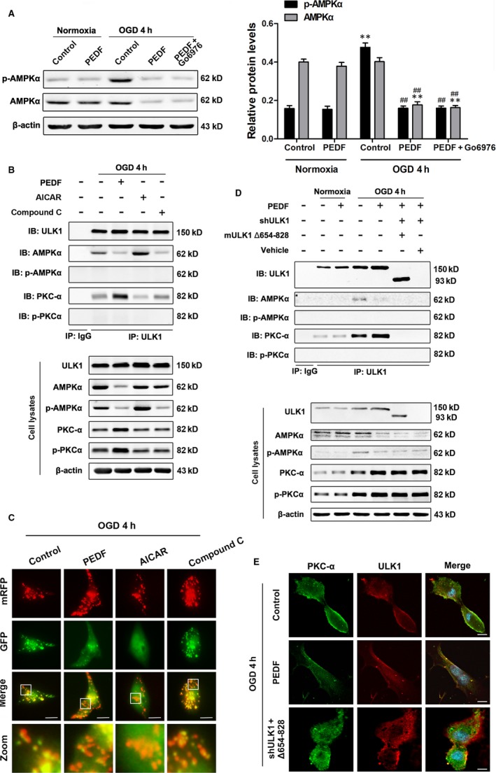
Figure 2.

PKCα interacts with the S/T domain of ULK1. A, Protein levels of p‐AMPKα, AMPKα in cardiomyocytes treated with PEDF or not under normal conditions or subjected to OGD for 4 hours, n = 6. B, Cardiomyocytes were treated with PEDF, AMPKα inhibitor (Compound C, 10 μmol/L) or AMPKα activator (AICAR, 1 mmol/L), then immunoprecipitated with ULK1 antibody. ULK1, AMPKα, p‐AMPKα, PKCα and p‐PKCα were determined using their antibodies, n = 4. IP: immunoprecipitation, IB: immunoblotting. C, Representative images showing LC3 staining in different groups of Cardiomyocytes infected with GFP‐RFP‐LC3 adenovirus for 24 hours, bar = 60 μm. Cardiomyocytes were treated with PEDF, AICAR and Compound C before OGD for 4 hours, n = 30 from three independent experiments. D, Cardiomyocytes were treated with PEDF, lentiviral short hairpin RNA targeting rat ULK1 (shULK1), deletion mutant ULK1 (mULK1 Δ654‐828) or empty vehicle, then immunoprecipitated with ULK1 antibody. ULK1, AMPKα, p‐AMPKα, PKCα and p‐PKCα were determined using their antibodies, n = 4. IP: immunoprecipitation, IB: immunoblotting. E, Confocal immunofluorescence analysis of PKCα interaction with ULK1 in cardiomyocytes, bar = 60 μm. **P < 0.01 vs normal control, ## P < 0.01 vs OGD control
