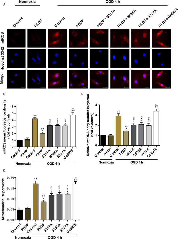
Figure 6.

The release of mitochondrial ROS and DNA in cytosol is decreased by PEDF‐induced phospho‐ULK1. A and B, Fluorescence microscope of (A) Mito‐SOX Red‐labelled ROS production and release in cytosol and quantification of (B) fluorescence density, bar = 100 μm, n = 30 from 3 independent experiments. Cardiomyocytes were treated with PEDF or PEDF+Go6976 under normal conditions or before OGD 4 hours. S317/555/777A mutants were transfected into cardiomyocytes as indicated. C, Cytosolic mitochondrial DNA copy number was measured by quantitative PCR, n = 7. Wild type or S317/555/777A mutational cardiomyocytes were treated with PEDF or Go6976 under normal conditions or before OGD 4 hours. D, Quantification of mitochondrial superoxide was detected with flow cytometric, n = 3. Wild type or S317/555/777A mutational cardiomyocytes were treated with PEDF or Go6976 under normal conditions or before OGD 4 hours. *P < 0.05, **P < 0.01 vs normal control, # P < 0.05, ## P < 0.01 vs OGD control, Δ P < 0.05, ΔΔ P < 0.01 vs OGD+PEDF group
