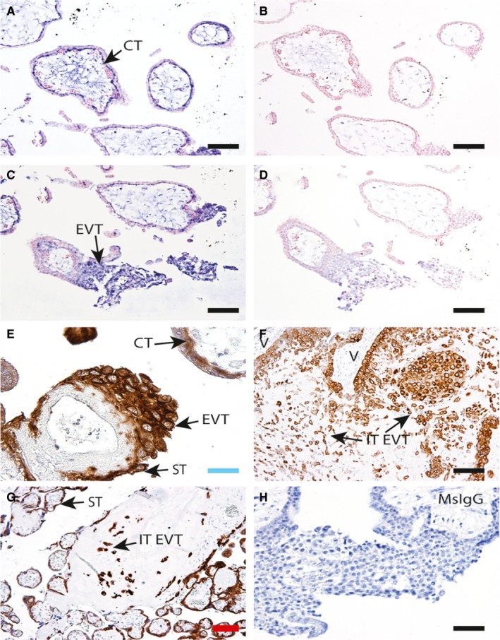
Figure 1.

ABCB1 mRNA and protein localization in CT and EVT. A and C, Representative images of first trimester placental sections showing specific ABCB1 antisense RNA (probe) hybridization to cytotrophoblast (CT) and extravillous trophoblast (EVT) columns (arrows). B and D, Sense controls in serial sections showing no staining. Representative immunohistochemistry (IHC) showing P‐gp protein localization in E, first trimester; F, 18w decidua basalis; and G, term placental bed and placenta. P‐gp staining is detected in syncytiotrophoblast (ST), CT and EVT in the first trimester and in interstitial EVT (IT‐EVT) within the decidua across gestation (arrows), (V = villous). H, Mouse IgG control. n = 4 placenta at each time‐point. Scale bars: blue = 25 μm, black = 50 μm, red = 100 μm
