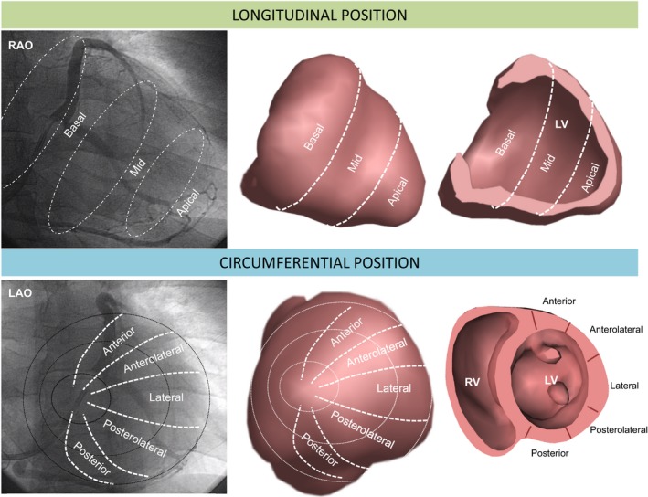
Figure 1.

Assessment of LV lead position using fluoroscopy. Longitudinal LV lead positions were assigned using the 30° RAO fluoroscopic view at the time of implantation, into basal, mid and apical. These correspond to the sectors shown in the 3‐dimensional long axis envelope and cross section (upper panel). Circumferential LV lead positions were assigned using the 30° LAO fluoroscopic view into anterior, anterolateral, lateral, posterolateral and posterior sectors, as shown in the short axis envelope and cross‐section (lower panel). LAO indicates left anterior oblique; LV, left ventricular; RV, right ventricular; RAO, right anterior oblique.
