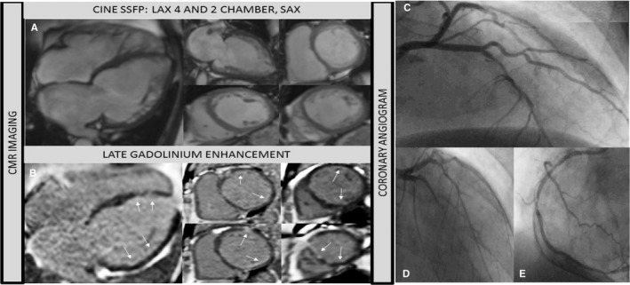
Figure 5.

Sixty‐five‐year‐old man. History of chronic heart disease and heart failure. Chest pain. No previous angiogram. CMR to assess viability. A, Cine steady free precession (4 chamber, 2 chamber and short axis views) showed bi‐ventricular dilatation and severe systolic dysfunction. B, Contrast CMR (LGE: 4 and short axis views) revealed sub‐endocardial enhancement in the ventricle (see arrows). CMR study suggests possible multi‐vessel CA disease with a low overall infarcted burden: global hibernation or dilated cardiomyopathy as differential diagnosis. Angiogram study (C through E) revealed triple vessel disease. Pending Coronary Artery Bypass Graft surgery. Angiogram images shared courtesy of Dr Milder Granados, Cayetano Heredia Hospital, Lima—Peru. CA indicates coronary artery; CMR, cardiac magnetic resonance; LGE, late gadolinium enhancement.
