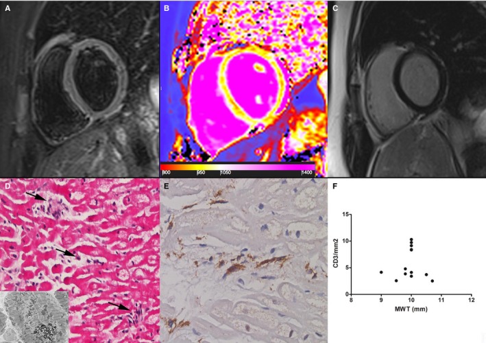
Figure 2.

Group 1 patient. A 22‐year‐old woman with prehypertrophic Fabry disease cardiomyopathy (maximal wall thickness [MWT]: 8 mm) showing normal myocardium in a T2‐weighted short τ inversion recovery image (A) and a late gadolinium–enhanced image (C). Analysis of T1 mapping (B) showed a reduced septal T1 value (923±30 ms, n.v. 960–990 ms), reflecting initial tissue lipid accumulation. D, At histology (hematoxylin and eosin, ×200), cardiomyocytes are intermittently enlarged because of cytoplasm vacuoles that, at electron microscopy, consist of myelin bodies (insert) and are focally (arrows) surrounded by CD3+ infiltrates (E) with cell necrosis. F, Correlation between MWT and CD3 count (P=ns) in all group 1 patients (n=13).
