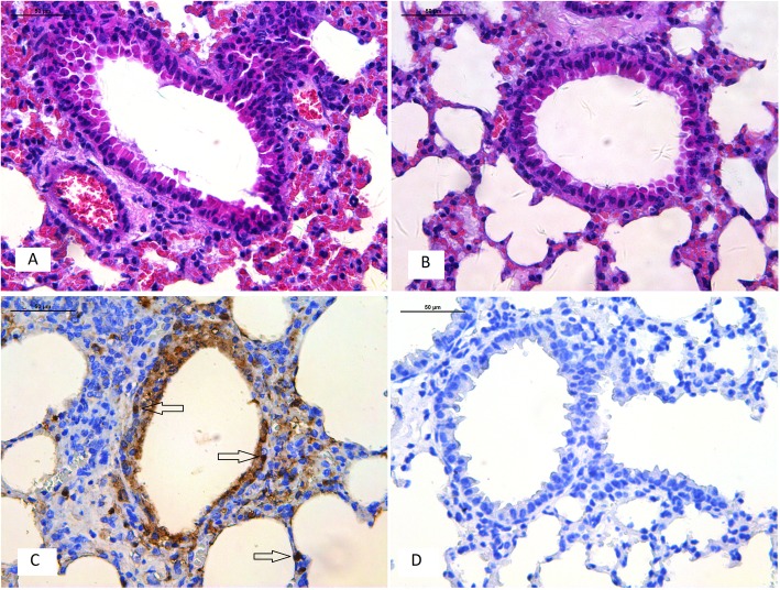
Fig. 5.
Histology and immunohistochemistry of mice infected with ZJ-1664 at 6 days post–infection. Histology of lung sections stained by hematoxylin and eosin from inoculated mice (a) or negative control (b). Immunohistochemical detection of virus nucleoprotein in lungs from inoculated mice (c) or negative control (d). Arrows indicate positively stained lung alveolar epithelial cells

