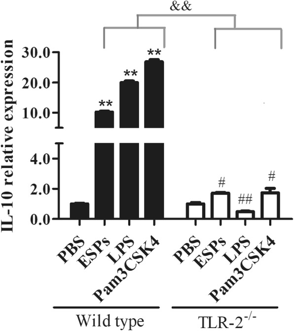
Fig. 3.

The comparison of IL-10 relative expression in wild type and TLR-2−/− B cells stimulated by EgPSC-ESPs. CD19+B cells isolated from wild type and TLR2−/− mice were cultured for 72 h in the presence of PBS, EgPSC-ESPs (5 μg/ml), LPS (10 μg/ml) or Pam3CSK4 (300 ng/ml). The relative expression of IL-10 in cultured B cells was compared. Data are expressed as means ± SD of triplicate wells in one round experiment (n = 6), and the results were repeated in three independent experiments. Differences were analyzed by one-way ANOVA or S-N-K method. VS PBS group in wild type B cells, **P < 0.001; VS PBS group in TLR-2−/− B cells, #P < 0.05; ##P < 0.001. &&P < 0.001, indicated the significant differences between EgPSC-ESPs, LPS and Pam3CSK4 (except for PBS) in wild type B cells and in TLR-2−/− B cells
