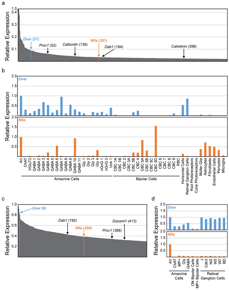
Figure 2: Transcriptome analysis of AII amacrine cells.
(a) Relative expression of the top 500 genes in adult AII amacrine cells, with known AII marker genes and new candidate marker genes highlighted with their relative position in parentheses (derived from Macosko et al., 2015 and Shekhar et al., 2016). (b) Expression of two candidate marker genes, Dner (blue top histogram) and Nfia (orange bottom histogram), across several cell populations, normalized to their expression in AII amacrine cells. (c) Relative expression of the top 400 genes in postnatal (P7) AII amacrine cells, with the position of several AII genes highlighted (derived from Kay et al., 2012). (d) Expression of Dner and Nfia in AII amacrine cells compared to other cell types/classes at P7.

