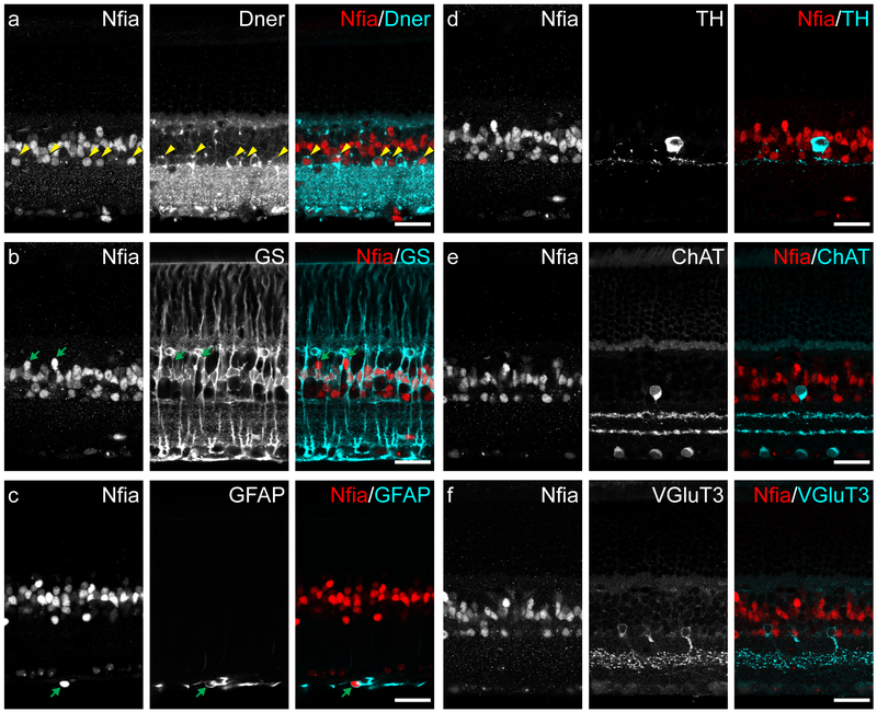
Figure 4: Localization of NFIA protein in adult retina.
(a) NFIA+ cell nuclei were found in the INL, both in the middle and along the inner edge. Many NFIA+ nuclei closest to the IPL (yellow arrowheads) were also DNER+, potentially indicative of the AII amacrine cell population. (b) Most NFIA+ nuclei in the middle of the INL were also GS+, and thus the population of Muller glia; the remaining NFIA+ profiles in this layer appeared to be a sparse type or types of bipolar cell (green arrows). (c) NFIA+ nuclei in the inner most edge of the retina, the nerve fiber layer, were surrounded by GFAP labeling (green arrow), indicative of being retinal astrocytes. (d) TH+ amacrine cells were not NFIA+. (e) ChAT+ amacrine cells were not NFIA+. (f) VGluT3+ amacrine cells were not NFIA+. Scale bars = 25 μm.

