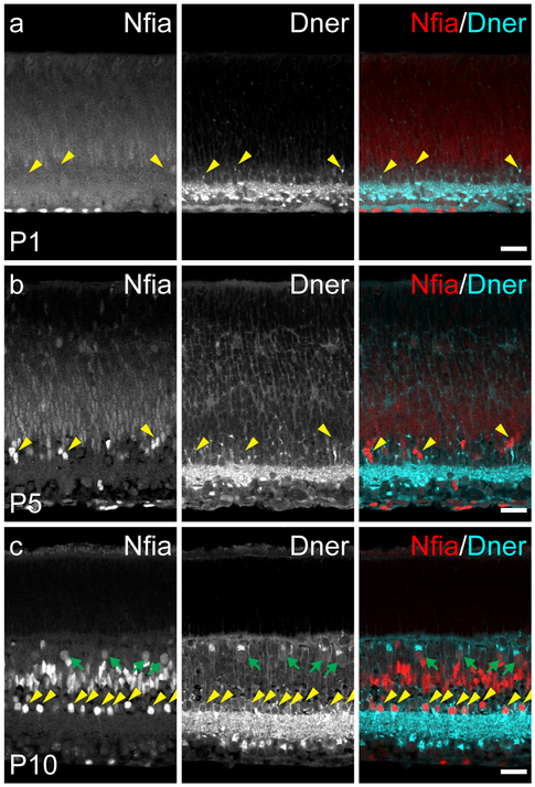
Figure 6: DNER and NFIA expression in postnatal retinal development.
(a) On the day of birth (P1), NFIA was only expressed in nuclei along the innermost margin of the retina, indicative of retinal astrocytes. DNER, by contrast, was expressed in the GCL and IPL, with some DNER+ stalks observed in the developing INL (yellow arrowheads). (b) At P5, NFIA was found in nuclei of cells in the INL, some of which also appeared to be juxtaposed to strong DNER labeling (yellow arrowheads). (c) At P10, NFIA was expressed in a population of amacrine cells along the inner margin of the INL, many cells in the middle of the INL, and a population of cells near the outer margin of the INL. DNER expression was maintained in the GCL and IPL, and now also expressed by cells in the amacrine cell division of the INL, identifying the population of AII amacrine cells by virtue of their coincident expression of NFIA (yellow arrowheads), and to the population of horizontal cells, which were also transiently NFIA+ at this age (green arrows). Scale bars = 25 μm.

