
Figure 8.
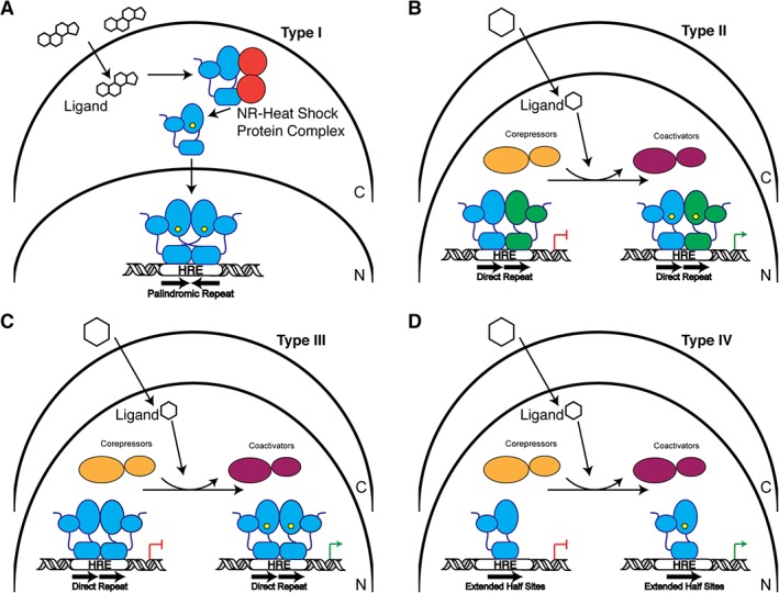
Schematic of NR signaling mechanisms. (A) Type I receptors reside in the cytoplasm (C) in complex with chaperone proteins. Upon ligand binding (hexagon), the receptor is released from this complex and is trafficked into the nucleus (N) where they typically bind to palindromic hormone response elements (HREs) as a homodimer to regulate transcription. (B) Type II receptors are localized in the nucleus. In their unliganded state, they interact with co‐repressor proteins, but upon ligand binding are exchanged for co‐activators. NRs in this group generally form heterodimeric complexes with RXR. (C) Similar to Type II receptors, Type III receptors reside in the nucleus and exchange bound co‐repressors and co‐activators. These receptors bind to direct repeat HREs as homodimers. (D) Type IV receptors are almost identical to Type III except they bind HREs that are extended half sites as monomers.