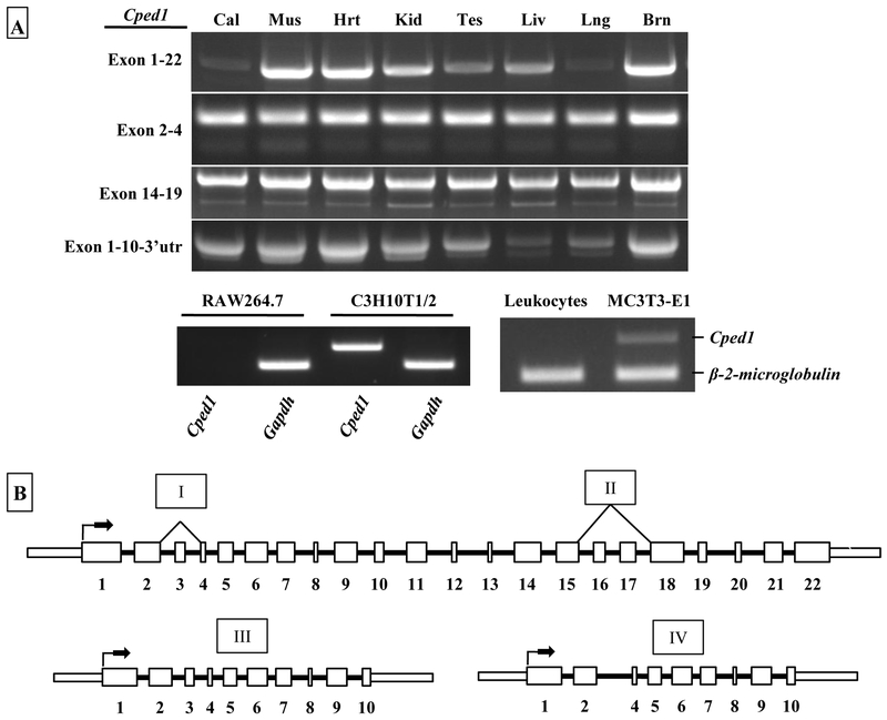
Figure 3: Cped1 isoforms are widely expressed in whole mouse organs but absent in RAW264.7 cells and circulating leukocytes.
A) RT-PCR for Cped1 transcripts was performed in eight C57BL/6J mouse tissues (Cal: calvaria, Mus: skeletal muscle [rectus femoris], Hrt: heart, Kid: kidney, Tes: testis, Liv: liver, Lng: lung, Brn: brain). Expression of a segment of exons 1 and 2 of Cped1 was tested in RAW264.7 monocytes/macrophases, multipotent C3H10T1/2 cells, MC3T3-E1 calvaria-derived pre-osteoblasts, and whole blood leukocytes. B) Summary schematic of alternative splicing suggests a diverse expression pattern for Cped1. Each of the identified and confirmed Cped1 transcripts including the splicing events (depicted above with roman numerals) is listed. Thick and thin rectangles represent exon and UTRs, respectively, and solid lines represent introns. Arrows indicate putative start sites for initiation of translation.

