Figure 1.
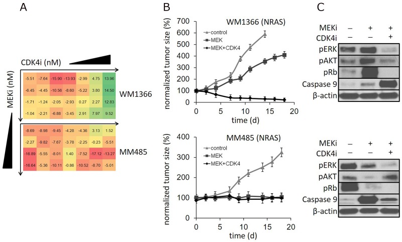
(A) NRAS mutant melanoma cell lines WM1366 and MM485 incubated with increasing concentrations of a MEK and CDK4,6 inhibitor in combination (MEKi: 1nM-125nM; CDK4,6i: 0.04nM-625nM). The numbers represent the relative change in viability compared to single MEK inhibitor treatment. (Color codes: linear range from ‘red’ - representing less reduction in cell viability by MEK/CDK4,6 compared to single MEK inhibition - to ‘green’ - representing increased reduction of cell viability by MEK/CDK4,6 compared to single MEK inhibition). (B) NRAS mutant human melanoma xenografts in mice treated with vehicle control, a MEK inhibitor or the MEK/CDK4,6 inhibitor combination: Tumor size reduction with MEK/CDK4,6 compared to single MEK inhibition of WM1366 tumors, but not of MM485 tumors. (C) Respective immunoblots of tumor tissue: Induction of p-Rb by single MEK inhibitor treatment in WM1366 tumors. In contrast, p-Rb reduction by single MEK inhibition in MM485 tumors. (*mice had to be euthanized due to tumor size, N=4).

