. 2018 Oct 25;7:e35854. doi: 10.7554/eLife.35854

Figure 4. tDCs intervention facilitates visual learning.

(a) Task performance (d') for the active stimulation groups (anodal, cathodal tDCS on posterior occipito-temporal cortex (Figure 4—figure supplement 1)) normalized to the sham group for the pre- and post- training blocks (no feedback, no stimulation) and the two training blocks (Block 1, Block 2; 500 trials per block). Performance (d') was significantly enhanced for anodal (but not cathodal) stimulation in the Signal-in-Noise task, while for cathodal (but not anodal) stimulation in the Feature differences task. (b). Behavioral improvement (d’ post- minus pre-training) was enhanced for the anodal stimulation group in the Signal-in-Noise task and the cathodal stimulation group for the Feature differences task. Error bars indicate standard error of the mean across participants.

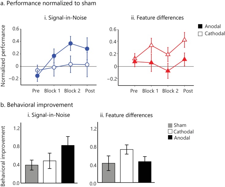
Figure 4—source data 1. Performance (d') normalized to the sham group for each participant of the anodal and cathodal groups for the two tasks.
DOI: 10.7554/eLife.35854.021

Figure 4.

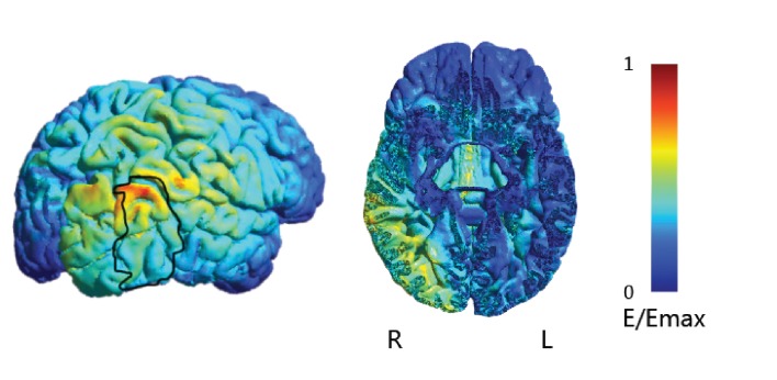
Figure 4—figure supplement 1. tDCs electric field simulation.

Figure 4—figure supplement 1.

Electrical field density simulation (shown on the cortical surface and a representative axial slice) showed that the current density was largely unilaterally localized, the peak of the electric field density was observed under the anode electrode around the posterior occipito-temporal cortex) and the stimulation reached the occipito-temporal region where the MRS voxel was placed. Heatmap indicates electric field strength from 0 (blue) to maximum (red). The black outline indicates mean activation across participants (n = 33) for an independent functional localizer scan (i.e. activation for intact vs. scrambled images of objects) that has been extensively used to identify regions in the posterior occipito-temporal cortex that are involved in shape processing (Kourtzi and Kanwisher, 2001). The figure illustrates substantial overlap between the tDCs electric field and regions in the posterior occipito-temporal cortex involved in shape processing. The exact mechanism by which tDCs alters cortical excitability remains debated: while some studies suggest that the electric field reaching the cortex in standard tDCs protocols is too weak to alter neuronal firing rate (Vöröslakos et al., 2018), other studies have shown that LFP power and coherence (Krause et al., 2017) are modulated with tDCs intensities between 1 and 2mA. Further, in vivo studies in humans have shown that lower current intensity of 1-2mA, similar to that used in our study, can induce voltage gradients that may still affect cells with an appropriate orientation relative to the applied field or already depolarized cells (Opitz et al., 2016; Widge, 2018).