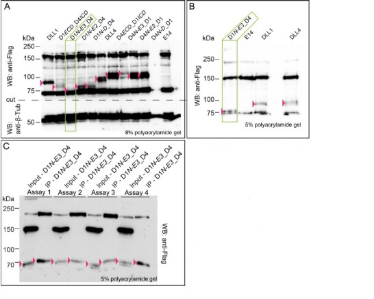
Author response image 1. Expression of D1N-E3_D4 cannot be quantified with confidence due to a closely co-migrating background band detected by the anti-Flag antibody.

(A) Expression of wild type DLL1 and DLL4 and of chimeric ligand proteins. Protein migration size varies due to differential post-translational modifications of the extracellular domains of DLL1 and DLL4. Double bands in DLL4, D4ECD_D1/CD, D4N-E3_D1, D4N-E2D1, and D4N-D_D1 lysates represent most likely differential modification states of the DLL4’s ECD. (B) D1N-E3_D4 migrates right at the dye front when using a 5% polyacrylamide gel. (C) Immunoprecipitation of D1N-E3_D4 after cell surface biotinylation in four independent assays. Input samples represents the whole cell lysate, IP samples represents the immunoprecipitated proteins. D1N-E3_D4 is detected in IP samples indicating its presence at the cell surface. The expression, however, cannot be quantified with confidence as the proteins are detected right at the dye front when using a 5% polyacrylamide gel. Pink arrowheads point to the proteins of interest. Green rectangles highlight the D1N-E3_D4 chimera lysate in (A) and (B).
