Figure 1.
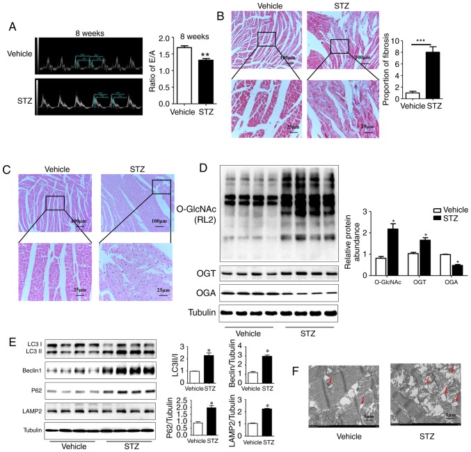
Myocardial injury is accompanied by an increase in O-GlcNAc modification and inhibited autophagic flux in rats at 8 weeks post-STZ induction. (A) M-mode echocardiography shows the left ventricular diastolic function as the ratio of E/A in rats at 8 weeks post-STZ induction. Morphological changes in the myocardium were assessed by (B) Masson staining and (C) hematoxylin and eosin staining (scale bar=100 and 25 µm). (D) Protein was extracted from heart tissue, and the expression levels of O-GlcNAc (RL2), OGT and OGA were detected by western blot analysis; (E) Expression levels of LC3II/I, LAMP2, Beclin-1 and P62 in each group were detected by western blot analysis in the vehicle group and the 8-week STZ-induced group. (F) Transmission electron microscopy shows autophagosomes, characterized by a double-layer membrane structure, as indicated with the red arrows (scale bar=1 µm). Data are expressed as the mean ± standard error of the mean (n≥6). *P<0.05 and **P<0.01, vs. Vehicle group. Tubulin was the loading control. O-GlcNAc, O-linked β-N-acetylglucosamine; STZ, streptozotocin; OGT, O-GlcNAc transferase; OGA, O-GlcNAcase; LAMP2, lysosome-associated membrane protein 2; LC3, microtubule-associated protein 1 light chain 3α; E/A, left ventricular filling peak velocity/atrial contraction flow peak velocity.

