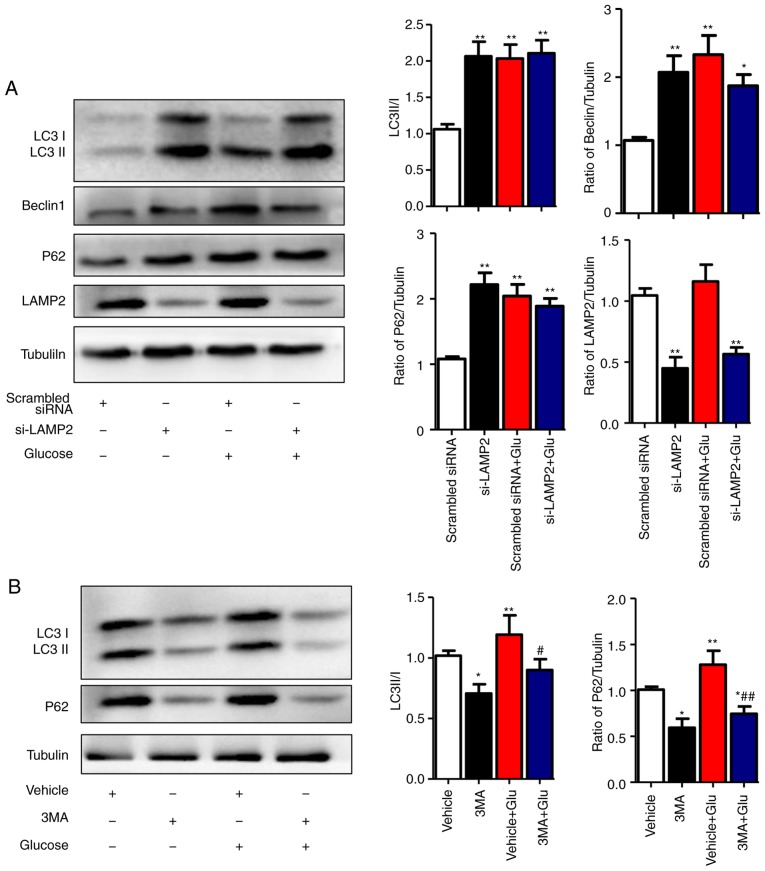
Figure 4.
High glucose inhibits the degradation stage of autophagy. (A) NRCMs were transfected with LAMP2 siRNA for 48 h and were exposed to high glucose for another 24 h. The expression of LAMP2, Beclin-1, P62, and LC3II/I in each group was detected by western blot analysis. (B) NRCMs were pretreated with 3-MA (5 mM) for 24 h and with high glucose for another 24 h; the expression levels of P62 and LC3II/I in each group were detected by western blot analysis. Data are expressed as the mean ± standard error of the mean (n≥6). *P<0.05 and **P<0.01, vs. Vehicle group; #P<0.05 and ##P<0.01, vs. Glu group. Tubulin was the loading control. NRCMs, neonatal rat cardiomyocytes; O-GlcNAc, O-linked β-N-acetylglucosamine; Glu, glucose; siRNA, small interfering RNA; LAMP2, lysosome-associated membrane protein 2; LC3, microtubule-associated protein 1 light chain 3α; 3-MA, 3-methyladenine.

