
Fig. 5. Giα2 acts independent of Rac1 activation in EGF-stimulated cell migration and it is required for actin polymerization and lamellipodia formation.
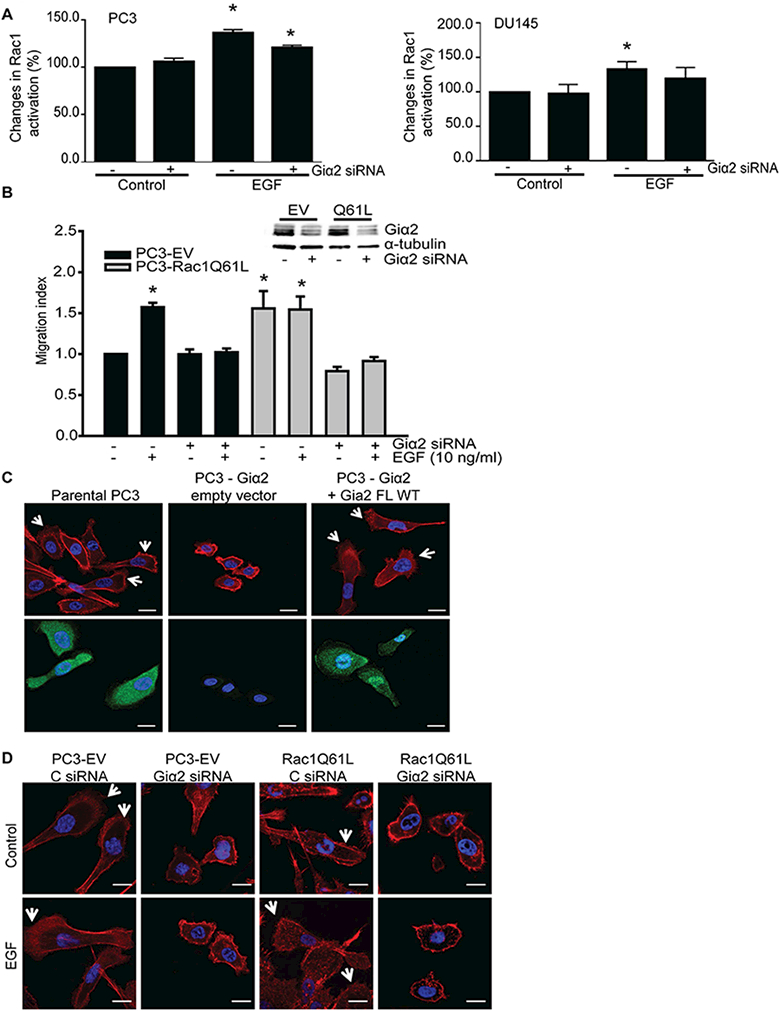
A. Activation of Rac1, after knockdown of endogenous Giα2. PC3 and DU145 cells were transfected with control siRNA or Giα2 siRNA for 48 hours; cells were serum starved overnight and stimulated with EGF (100 ng/ml) for 3 minutes and Rac1 activity was then measured with G-LISA kit. Data were expressed as mean ± SEM (n=3) and analyzed by ANOVA and Duncan’s modified range tests. * Significantly different (P<0.05) compared to controls. B. Upper panel shows Western blot analysis of Giα2 in cell lysates, to validate the knockdown of the protein. Cell migration in parental PC3-EV and PC3-Rac1Q61L cells was performed after knockdown of endogenous Giα2, in response to EGF (10 ng/ml). Results are expressed as migration index. Each bar represents mean ± SEM (n=3). * Significantly different (P<0.05) compared to controls. C. F-actin staining (red color, upper panel) and immunofluorescence (green color, lower panel) of Giα2, in parental PC3, PC3-Giα2 and PC3-Giα2+Giα2 FL WT cells. The arrows indicate the lamellipodia structures present in normal PC3 and in the restored PC3-Giα2+Giα2 WT. Cells were visualized with 40× objective. The scale bars represent 20 μm. D. PC3-EV cells and PC3-Rac1Q61L cells were transfected with control siRNA and Giα2 siRNA and incubated without (control) or with EGF (10 ng/ml) for 30 minutes. Cells were stained for F-actin (red) and DAPI (blue). Arrows indicate lamellipodia at cell edges. Cells were visualized with 40× objective. The scale = 20 μm.