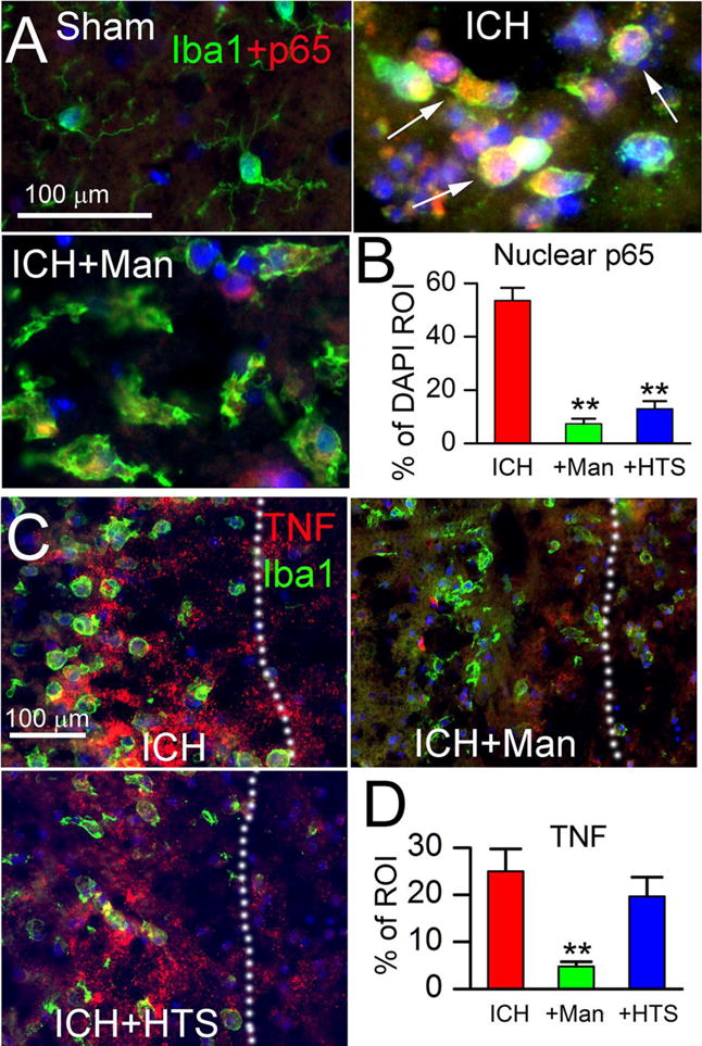
Figure 3. Mannitol and hypertonic saline attenuate p65 and TNF.

A,B: Double immunolabeling (merged images) at 48 hours for Iba1 (green) and p65 (red), with nuclear staining by DAPI, ipsilateral to the ICH (moderate-dose collagenase model), for sham injury, ICH without treatment (ICH), and ICH with mannitol treatment (ICH+Man), as indicated; arrows point to p65+ (red) and DAPI+ (blue) nuclei that appear pink; the bar graphs show quantification of p65 within DAPI+ nuclei (% of DAPI ROI) in the three conditions indicated (B); **, P<0.01 for treatments compared to no treatment; n=5/group. C,D: Double immunolabeling (merged images) at 48 hours for Iba1 (green) and TNF (red), ipsilateral to the ICH (moderate-dose collagenase model), for ICH without treatment (ICH), ICH with mannitol treatment (ICH+Man), and ICH with hypertonic saline treatment (ICH+HTS), as indicated; the dotted line demarcates the hematoma from perihematomal tissues; the bar graphs show quantification of TNF (% of ROI) in the three conditions indicated (D); **, P<0.01 for treatments compared to no treatment; n=5/group.
