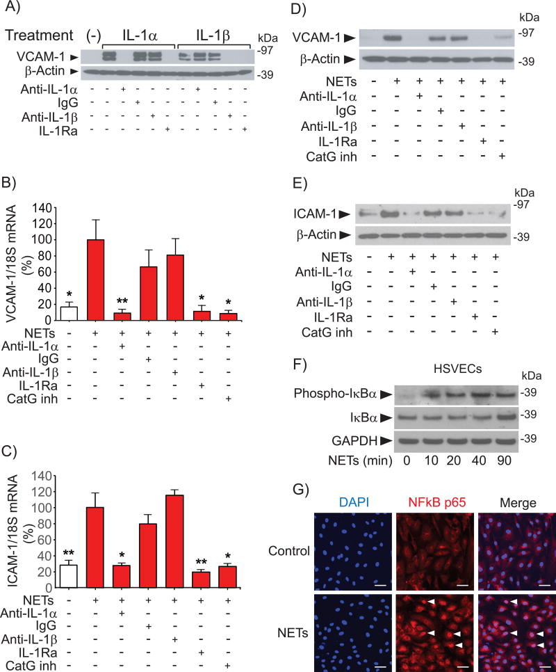
Figure 2. IL-1α and CatG mediate NET-induced expression of leukocyte adhesion molecules in HSVECs.
(A) Cells were incubated with 50 pg/ml IL-1α or IL-1β for 3 hours in the presence of the indicated antibodies (20 ng/ml) or IL-1Ra (1 µg/ml). Whole-cell extracts were fractionated by SDS-PAGE and immunoblotted with antibodies to VCAM-1 or β-actin. (B–C) Cells were incubated with NETs (0.5 µg DNA/ml) for 3 hours in the presence of the indicated antibodies (20 ng/ml), IL-1Ra (1 µg/ml), or CatG inhibitor I (50 µM), followed by RNA extraction and determination of VCAM-1 and ICAM-1 mRNA levels by RT-qPCR. N = 3–7. P values: *<0.05, **<0.01 vs. NETs alone, defined as 100%. (D–E) Cells were incubated as in (B-C) for 6 hours, followed by fractionation of whole-cell extracts by SDS-PAGE and immunoblotting with antibodies to VCAM-1, ICAM-1, or β-actin. (F) Cells were incubated with NETs (0.5 µg DNA/ml) for the indicated periods of time, followed by fractionation of whole-cell extracts by SDS-PAGE and immunoblotting with antibodies to phospho-IκBa, IκBα, or GAPDH. (G) Cells were incubated with NETs (0.5 µg DNA/ml) for 90 minutes, fixed, and stained with anti-NFκB p65 (1/1000 dilution) and DAPI as described in the Materials and Methods section. The arrowheads indicate NFκB localized to the nucleus. Scale bar = 50 µM. Omission of the primary antibody yielded no signal.

